
"Parcellating cortical functional networks in individuals"
Nat. Neurosci, 2015

"Individual variability in functional connectivity architecture of the human brain"
Neuron, 2013

"Cerebellar asymmetry and its relation to cerebral asymmetry estimated by intrinsic functional connectivity"
J Neurophysiol, 2013
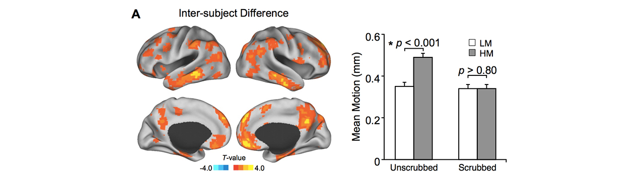
"Neurobiological basis of head motion in brain imaging"
PNAS, 2014
No two individuals are alike, however, the extent to which brain networks can differ between people is often underestimated in many clinical practices. We systematically investigated the inter-individual variability in functional organization, and found that it is the higher cognitive regions that are most variable between subjects. These areas are known to be activated when the brain performs complex tasks, and when we feel emotions. Besides providing better knowledge of how our brain works, this research has clinical importance, since knowing the individual variability in functional organization is crucial for the development of customized therapeutic approaches. When interventions (surgery, brain stimulation) are provided in areas of high variability, doctors could perform specific tests to check the relevance of standardized procedures. Building upon these findings, we recently developed a novel technology that can accurately map the unique functional network organization in each individual subject. This will benefit patients suffering from neurological and psychiatric diseases by providing critical information to determine the optimal treatment strategy for each individual, and to gain a deep understanding of the therapeutic effect in a particular patient.
It is well established that the human brain possesses a number of systems that are specialized between the two hemispheres. Past theories of lateralization have assumed a single factor that controls brain lateralization. The upshot has been a 40-year quest for an explanatory factor that controls the development of brain asymmetries. Recently, we developed a general data-driven approach to map the specialization of brain systems. Our study suggested that the single-factor models are incomplete and replaced them with a new multiple-factor model based on strong evidence that different mechanisms control lateralization for distinct brain systems. Further, we found a gradient of specialization across different functional regions and discovered that one specific association network, called the frontoparietal network, possesses a unique pattern that may suggest how specialization may emerge in the human brain. What we discovered is that the frontoparietal network is differentially coupled to distinct systems in each hemisphere. Thus, differentiation between the hemispheres may arise by how networks interact with distinct partner networks in each of the hemispheres. This discovery and the novel approach developed during the study were then immediately translated to clinical research. In a group of patients with schizophrenia, we found aberrant hemispheric specialization in the caudate nucleus that may relate to the development of illness.
We have been refining and validating the functional connectivity MRI technology in clinical populations. In 2009, we first demonstrated the feasibility of mapping functional networks in epilepsy surgical candidates based on functional connectivity. Our recent work showed that spontaneous and task-based mapping can be performed together using the same pre-operative fMRI data, provide complimentary information relevant for functional localization, and can be combined to improve identification of eloquent cortex in epilepsy surgical candidates. Additionally, we have been validating functional connectivity MRI technology in patients with brain lesions and demonstrated that functional connectivity MRI is very sensitive to the integrity of specific functional circuits.
An important direction in clinical neurophysiology research is to estimate the neural sources that give rise to the electrical activity (EEG) as well as the magnetic fields (MEG) measured on scalp. We developed a series of new algorithms to determine the locations and time courses of neural activity within the brain under various disease conditions. These algorithms are used in many hospitals to assist daily diagnosis. We also designed novel signal processing method to analyze intracranial EEG recorded from surgical patients and studied the spatiotemporal procedure of object recognition in human visual cortex.