Research Technology
We currently use structural magnetic resonance imaging (MRI), functional magnetic resonance imaging (fMRI), diffusion tensor imaging (DTI), and magnetoencephalography (MEG) to study the functional and structural determinants of movement recovery after stroke. We have also developed a set of sensors for measuring hand motion during imaging studies.
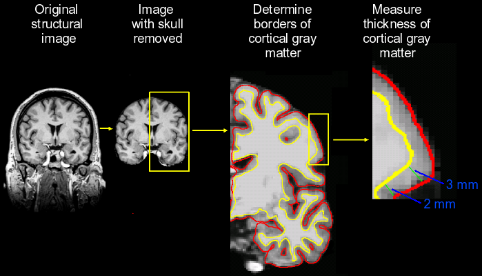
Structural Magnetic Resonance Imaging (MRI)
Magnetic resonance imaging can be used to obtain high-resolution images of the anatomy of the brain. We use these images to build a model of each subject's cortex and combine these cortical models together with images of brain activity (see fMRI) to localize sites of activity. In our laboratory, we also use structural images to measure the thickness of the cortex. Cortical thickness can change in response to stroke or repeated exposure to particular experiences.
|
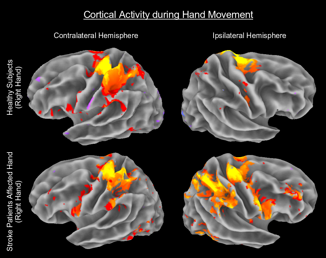
Functional Magnetic Resonance Imaging (fMRI)
Functional MRI is a type of MRI that allows us to examine changes in blood flow to local parts of brain, which provides an indirect measure of brain activity. Changes in brain activity occur when a subject performs a movement or responds to stimulation. The level and pattern of activity can be altered in a person with a brain injury such as stroke or in response to therapies that improve motor function. One of our goals is to better characterize the relationship between changes in brain activity and recovery of movement after stroke.
|
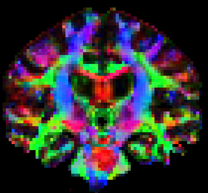
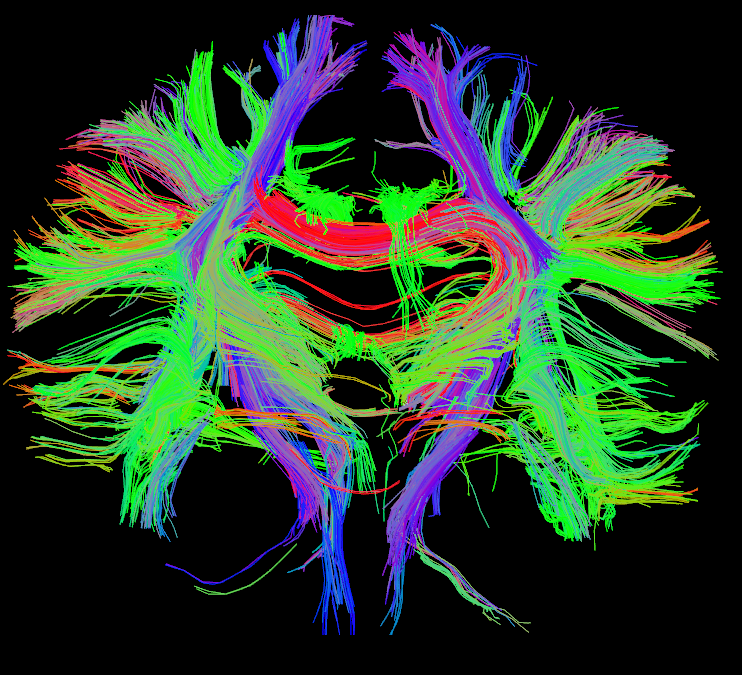
Diffusion Tensor Imaging (DTI)
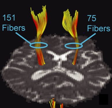
Diffusion tensor imaging is a type of MRI that allows us to measure microscopic movement of water in the brain. DTI can be used to evaluate the integrity of the white matter in the brain. White matter allows signals from one region of brain to be transferred to another region of brain. One particularly important white matter pathway for our research is the corticospinal tract. This tract allows the brain to send signals to muscle to control movement. Damage to the corticospinal tract causes loss of movement ability. The rightmost image below shows the effect of stroke on the corticospinal tract, measured by fewer fibers on the side of the stroke than on the other side.
|
|
|
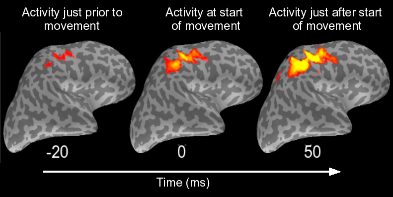
Magnetoencephalography (MEG)
Magnetoencephalography is a method for examining brain activity by measuring the minute magnetic fields on the scalp of a subject that are created by electrical activity of neurons in the brain. A major benefit of MEG compared to other methods of monitoring brain activity is its very high temporal resolution, allowing measurement of brain activity every millisecond or so. The data collected using MEG can be analyzed in a number of ways. One important method is analyzing brain activity across a range of frequencies. As shown below, the characteristics of brain activity change over frequency and over time relative to hand movement.
|
|

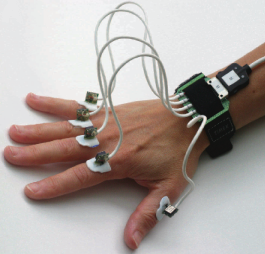
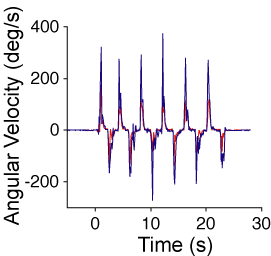
Finger Motion Sensors
In addition to these technologies, it is also important for us to characterize hand motion during imaging. To do this, we use a custom-built set of finger motion sensors that allow us to measure, with high-sensitivity, the speed at which each of the fingers moves.
|
|