
Research
Advanced Fluorescence Molecular Imaging Technologies for Alzheimer’s disease
Compared to MRI and PET, fluorescence imaging is much cheaper and suitable for high throughput screening. Fluorescence molecular imaging has been widely used as a tool for basic research and preclinical studies because of its cost-efficiency and high sensitivity. For small animal imaging, fluorescence molecular imaging could be acquired through conventional intensity-based imaging. However, to achieve more accurate information about the target, advanced technologies are necessary.
For intensity-based NIR imaging, in the absence of a “ smart” probe, in vivo differentiation between wild type and transgenic mice always depends on the retention of the probe, which requires waiting time. However, the probe with a “smart” property (such as intensity increase upon interacting with the target) could be used to differentiate wild type and transgenic mice at the very early time points after injection. Yet the signals from these intensity-based images cannot reflect the real time concentration of the target, since it represents the sum signal from both bound and free probe. To differentiate bound probe from free probe, two advanced imaging technologies could be relied on if the probe is a “smart” probe.
Spectral Unmixing Technology
Spectral unmixing technique is a wavelength-based imaging. When this technique was coupled with a “smart” probe, which displays a significant emission wavelength shift upon binding to a target, it will enable us to differentiate bound and unbound probe based on their spectra. Therefore, the signal from the bound probe in this case reflects the concentration of the target in real time if the “smart” probe saturates the target. In other word, the quantification of the target content will not be dependent on the injected dosages if the target is saturated. We believe this technology is able to report the Ab content in real time once the Ab was saturated.
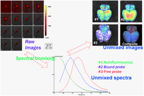
Picture 5: Spectral unmixing imaging
Lifetime-resolved imaging
Each fluorophore has its fluorescence lifetime. When a fluorophore interacts with its target, its lifetime may change. For a “smart” probe, it may have two lifetimes corresponding to bound form and free form. Similarly, as with spectral unmixing technology, lifetime-resolved imaging is able to give more accurate information regarding the content of the target, and the quantification is not correlated with the injection doses if the target is saturated.
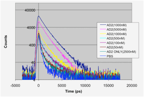
Picture 6: Lifetime imaging