
Research
Multimodal imaging of Alzheimer’s disease
Multi-modality imaging, an emerging field, has the capability to provide anatomical, functional, and molecular information by combining information acquired with different modalities of the same target. To fully take advantage of multi-modality imaging techniques, development of multi-modal contrast probes is highly desirable.
To create a multi-functional probe, a flexible scaffold is a necessary element. Such a scaffold could be composed of large assemblies such as nanoparticles, or alternatively, small molecules. Nanoparticles are ideal scaffolds for multi-modal probes because they have the flexibility to be modifiable with different MRI, NIR, Raman, PET or SPECT imaging tags. Currently, major efforts are focused on developing nanoparticles as multi-modal carriers for the imaging of cancer, stroke, and cardiovascular diseases. However, the development of a small molecule scaffold for a multi-modal probe is more challenging and has been less explored, possibly due to the poor flexibility and related modifiability of small molecules. Despite the difficulty of modification, exploring small molecular scaffold-based multi-modal probes will be tremendously useful for multi-modality imaging of neurodegenerative diseases, such as Alzheimer’s Disease, Parkinson’s Disease, and Huntington’s Disease.
In our studies, we propose to start with the development of a preclinical useful NIR (near-infrared) probe, to which we will make minor structural modifications to produce a clinically applicable multi-modal MRI and PET probe. We hope this approach will provide the means for future translation from preclinical research to clinical study.
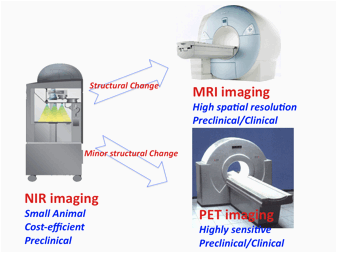
Picture 4: Flow chart from NIR to PET/MRI