

The Daily Free Press recounts the HUBWeek event in which Center Director Bruce Rosen and medical illustrator Danny Quirk spoke about the intersectionality of human anatomy and visual art.
Buckle Up: With New Techniques, MRI Is Faster Than Ever Before
July 14, 2015
 |
|
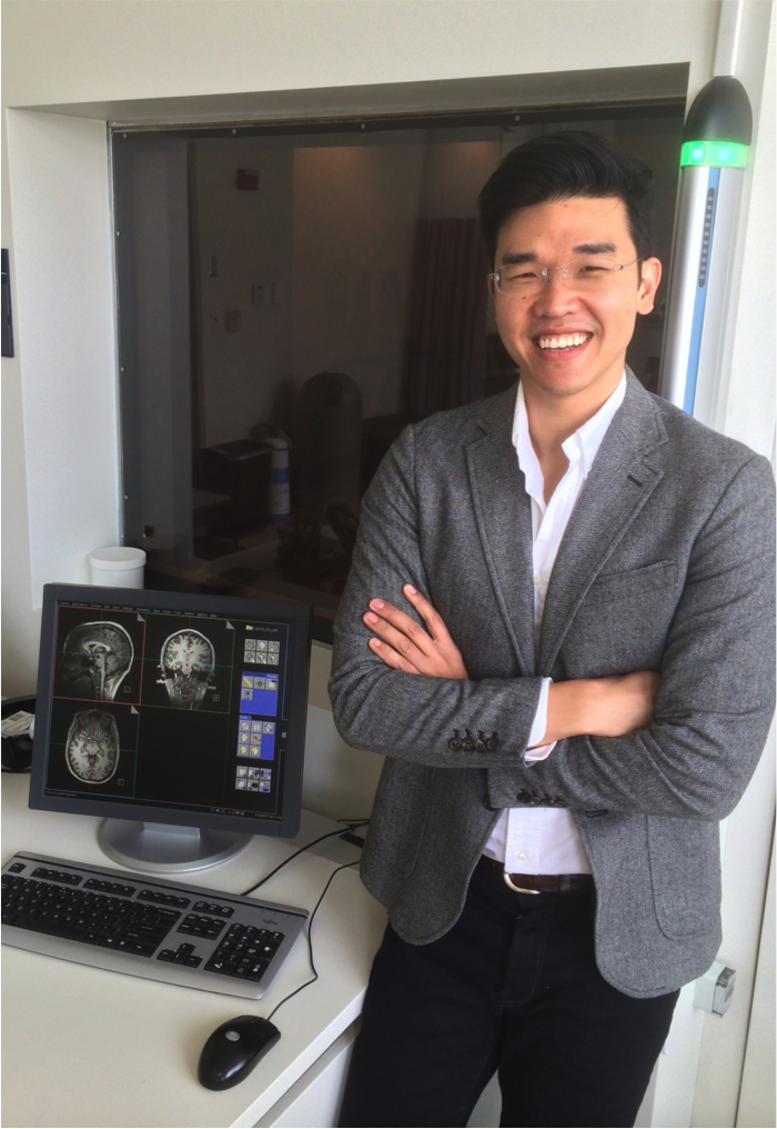
The Martinos Center’s Kawin Setsompop is developing advanced techniques that faciliate dramatic increases in MR imaging speed
|
With its exquisite soft-tissue contrast and unique ability to probe brain function, magnetic resonance imaging (MRI) has revolutionized our understanding of the brain in both health and disease. But because of its speed—it acquires scans at a relatively slow clip—it doesn’t always meet the needs of today’s cutting-edge applications.
Kawin Setsompop is looking to change this.
Setsompop, an investigator in the MGH Martinos Center for Biomedical Imaging and an Assistant Professor at Harvard Medical School, is developing new techniques for MRI that effectively speed up scans, thus yielding dramatically higher imaging resolution. By optimizing the interplay between imaging hardware, MR physics and neuroscience, the techniques enable study of the living, functioning brain at much finer scales than was previously possible. Setsompop says his goal is to increase the sensitivity and efficiency of MRI by an order of magnitude or more.
Already, the advances he’s described could have a major impact on healthcare. Implementing them will improve detection of subtle changes in both structure and function in the brain, and this in turn will benefit a range of applications. Being able to identify such changes can play a vital role in, for example, the diagnosis, prognosis and treatment of central nervous system (CNS) disorders including multiple sclerosis and epilepsy.
Setsompop’s interest in helping people by advancing biomedical imaging technology dates back to the middle of the last decade, to his graduate school days at MIT studying electrical engineering and computer science. In deciding on a PhD project, he found he was drawn to research in the health sciences. He knew, he says, that this is where his training would have the greatest impact on society.
Working with Elfar Adalsteinsson, director of the Magnetic Resonance Imaging Group at MIT, he started tackling a problem associated with a technique known as ultra-high-field MRI. A relatively new advance at the time, ultra-high-field MRI suffered from an inherent limitation: the higher field strengths used with the technology led to inhomogeneity in the magnetic field, and this produced artifacts in the image. Hoping to address this—and thus to improve the overall efficacy of the approach—Setsompop and Adalsteinsson and colleagues started developing strategies that could mitigate these effects.
He continued his efforts after joining the Martinos Center as a postdoctoral fellow working with Larry Wald. In the labs and conference rooms of the Center’s campus in the historic Charlestown Navy Yard in Boston, he complemented his signal processing and algorithm design training with additional training in MRI physics, hardware development and neuroscience—training received in Dr. Wald’s group but also informally through interactions with researchers across the Center. This proved an integral part of his growth as an investigator, he says, and an experience that could really only be had in an environment like the Center, a crossroads of research and thought in a broad range of biomedical imaging-related fields.
He found the interdisciplinary nature of the work tremendously appealing. At the same time, he was captivated by just how quickly a new idea can become a workable prototype. “In this research field, an innovative idea can be developed, tested and demonstrated within a few days,” he says, and if successful can have a significant impact in both the neuro-scientific and medical communities. “This was eye-opening for me and it still makes me excited every day, thinking about and tinkering with new ideas.”
Among the many fruitful ideas he’s had: the new strategies to speed up MRI scans. While still a postdoctoral fellow, Setsompop introduced the technique known as blipped-CAIPI Simultaneous MultiSlice (SMS) imaging. This allows investigators to acquire up to ten planes, or “slices,” of brain images at a time—instead of just one—enabling much faster snapshots of brain physiology. Researchers around the world immediately saw the advantages of this approach and in the few short years since a robust community of users has taken shape. Recognizing this, Setsompop and David Feinberg of the University of California, Berkeley, recently put together a retrospective virtual issue of the high-impact journal Magnetic Resonance in Medicine highlighting progress in the field. This coincides with a workshop that he and Feinberg and others are organizing—the very first ISMRM-sponsored workshop on the technique: “Simultaneous Multi-Slice imaging: Neuroscience and Clinical Applications”—to be held in Pacific Grove, Calif., July 19-22, 2015.
To understand the potential impact of the technique, we need look no farther than two recent high-profile initiatives. First, researchers have used blipped-CAIPI SMS imaging to acquire all of the fMRI and diffusion data in the large-scale NIH-funded Human Connectome Project. The HCP encompasses two collaborations, one between the MGH Martinos Center and the Laboratory of Neuro Imaging at the University of Southern California (see here), the other between groups at Washington University and the University of Minnesota. WIth both of these, the investigators have laid out the goal of constructing a complete map of the structural and functional neural connections in humans, in vivo. Blipped-CAIPI SMS imaging is providing the unprecedented resolution in MR imaging that is needed to achieve this.
Also, the technique will play an integral role in a multi-institutional project supported by the White House’s BRAIN Initiative; the $1.4 million award was among the first wave of grants to be funded by the National Institutes of Health as part of the initiative, which is designed to advance understandings of the human brain through innovative neurotechnologies. The cross-institutional team is now working to develop a new technique—MR Corticography (MRCoG) or, simply, cortical MRI—that will increase the level of detail in seen brain scans by more than 30 times over today’s most powerful MRI systems. Using this, they hope to see features as small as 200 microns across, or about twice the width of a human hair.
Through these and other research projects, blipped-CAIPI SMS imaging is furthering—in often dramatic ways—our understandings of how the brain works. Importantly, Setsompop’s efforts can also help to advance a range of clinical applications.
Just one example: He and his team are currently fine-tuning the next generation of simultaneous multislice imaging techniques. Known as Wave-CAIPI SMS, this will provide an order of magnitude improvement in data acquisition efficiency for a variety of clinically important MRI scans. Once it’s ready to go, he says, this technology will enable clinicians either to speed up current MRI clinical exams—ultimately leading to significant reductions in healthcare costs—or to obtain much more detailed scans in the same time it takes to complete a typical clinical exam today.
-----
Subscribe to the Martinos Center newsletter to read more about what's happening in the Center.