

 Transcranial Magnetic Stimulation (TMS) is a non-invasive technique used to stimulate the human brain. It was invented in 1985 and has since been used extensively for research and clinical purposes.
When you see a picture, hear a sound, or think of something, electric currents driven by electric fields will flow inside certain parts of your brain. TMS causes similar electric currents in the brain, but the neurons are instead activated with a magnetic pulse from a coil that is placed over the head. In TMS, the electric fields are generated by electromagnetic induction. Therefore, they travel effortlessly through the scalp and skull, which makes TMS an easy, painless, and non-invasive way to stimulate the brain.
TMS has a unique role in understanding how the brain works. Brain imaging techniques such as electroencephalography (EEG), magnetoencephalography (MEG), and functional magnetic resonance imaging (fMRI) record brain activity and can tell us where and when the activations occurred. However, they cannot tell if a specific activation is necessary for a given task. TMS can be used for turning a specific brain area "off" for a small fraction of as second. Therefore, TMS allows establishing causality between brain activations and different types of sensory, motor, and cognitive functions.
TMS may also be useful for diagnosis and treatment of some clinical conditions, such as migraine headache and depression, and for pre-surgical mapping of motor functions.
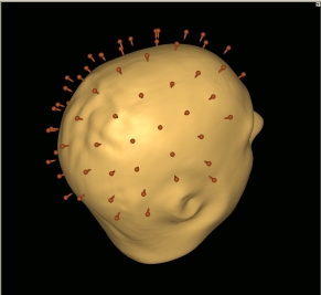
The effects of TMS depend on which brain area is stimulated. Therefore, TMS is often combined with navigation devices that allow accurate localization of the stimulated area. TMS is also typically done with electromyogram (EMG) that measures electrical activity of muscles, and behavioral measurements such as reaction times. Finally, to observe the effects of TMS directly on the brain we may also record EEG, fMRI, or PET simultaneously with TMS. Such multi-modal combinations of techniques are powerful in studying how the brain works in health and in disease.
Our TMS laboratory is equipped with two MagPro X100 with MagOption stimulators, multiple MagPro (including MRI-compatible and liquid-cooled) coils, a Nexstim eXimia Navigated Brain Stimulation (NBS) frameless neuronavigator integrated with a 6-channel EMG unit, and a Nexstim eXimia 60-channel EEG system. To ensure exceptionally clean EEG recordings, the instruments are located within an electrically shielded room. Computer-controlled visual, auditory, and somatosensory stimulation systems as well as behavioral response monitoring are available in the laboratory. Our analysis software allows integration of TMS, EEG, MEG, anatomical MRI, and fMRI data. The TMS laboratory is located inside the Biomedical Imaging Core (BIC) at the Martinos Center that is ideally equipped to support clinical and pharmacological studies and experiments that require advanced physiological monitoring.
You can learn more about TMS here. You may also be interested in seeing the TMS Wiki page here.