

The Daily Free Press recounts the HUBWeek event in which Center Director Bruce Rosen and medical illustrator Danny Quirk spoke about the intersectionality of human anatomy and visual art.
The Boston Globe talks to Bruce Rosen about the history of MRI
October 14, 2014
This week's issue of the Boston Globe Magazine is devoted to the human brain. So it only makes sense that its editors turned to Bruce Rosen, director of the MGH Martinos Center for Biomedical Imaging, to talk about brain imaging. In "This Is Your Brain on MRIs," Dr. Rosen walks them through the history of MRI-based imaging techniques: from early MRI scanners installed at MGH to functional MRI—which was pioneered at the Martinos Center in the 1990s—and on again to Connectome imaging, the purpose of which is "nothing less than mapping the myriad neural connections in the brain that sustain consciousness," says the Globe.
The Martinos Center is recognized worldwide as a leader in the development and application of imaging strategies for biomedical research and care. These include cutting-edge imaging techniques—positron emission tomography, MEG/EEG, optical imaging and more, as well as the MRI-based methods noted above—offering unique insights in the brain and elsewhere the body.
The Globe article is online here.
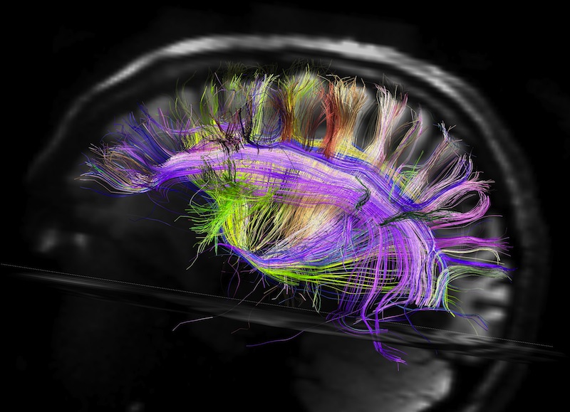
This image, obtained by the MGH Martinos Center's Van Wedeen and Larry Wald as part of the Human Connectome Project, illustrates the recent discovery that brain pathways are organized geometrically, in 2D sheets and 3D grids.