

The Daily Free Press recounts the HUBWeek event in which Center Director Bruce Rosen and medical illustrator Danny Quirk spoke about the intersectionality of human anatomy and visual art.
New Technique Paves The Way For Clinical Application Of fMRI
November 9, 2015
 |
|
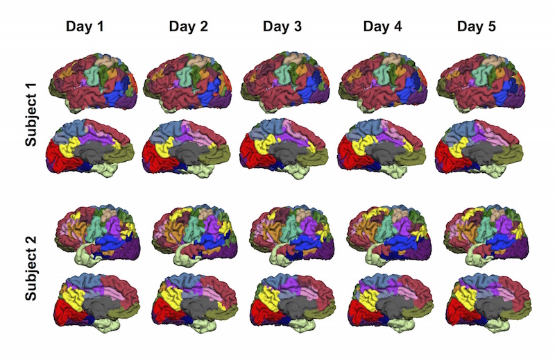
Researchers have reported a technique for mapping functional brain areas in individual subjects, opening the door to a range of clinical applications of functional MRI. Using the technique, they delineated the subjects’ brains into 18 different networks. The images here show the variability across subjects—especially in higher-order association areas like the language network in the frontal lobe—as well as the reproducibility within subjects over time.
|
Functional MRI has revolutionized our understandings of how the brain works, offering ever deeper insights into the neural underpinnings of a range of behaviors. But because it typically relies on averaged findings from groups of subjects, it isn’t yet established in the clinic, where the focus is necessarily on the individual.
Now, the Martinos Center’s Danhong Wang and Hesheng Liu and colleagues have reported a technique that could usher in a new era of clinical fMRI by providing a unique, personalized map of the different functional areas of an individual’s brain. They describe the technique in a Nature Neuroscience paper published online today.
“There is an urgent need to develop individual-based neuroimaging technologies to guide and facilitate personalized medicine,” said Liu, an Assistant Professor of Radiology at Harvard Medical School and Director of the Laboratory for the Study of the Brain Basis of Individual Differences at the MGH Martinos Center for Biomedical Imaging. “By identifying the functional architecture of the brain at the individual level, our new fMRI-based technique could advance clinical applications including neurosurgical planning and treatment and management of a range of neurological and psychiatric disorders.”
The cerebral cortex in humans is organized into distinct areas, each of which is responsible for a specialized function that plays a role in a larger, distributed network. We know, however, that there are differences between individuals in how these areas are organized. If we are to fully understand the relationship between anatomy and function, and particularly if we are to apply this understanding to any of a range of clinical needs, we need to be able to discern—precisely and reliably—what those differences are.
To this end, the authors of the Nature Neuroscience paper developed and tested a “cortical parcellation” approach that can be used with either resting-state fMRI data or spontaneous activity extracted from task-based fMRI data. This novel method begins with a cortical atlas derived from data from 1,000 healthy subjects, Liu said, and iteratively finds the corresponding atlas in the individual subject by correlating fMRI signals from each vertex with atlas-based reference signals. The procedure incorporates a priori knowledge such as the distribution of inter-subject variability in the brain, which was estimated by Liu’s lab in a recent Neuron paper.
In a series of tests, the functional maps produced by the new approach proved highly reproducible while also capturing variability across subjects. The researchers validated these results using invasive mapping methods in surgical patients.
All of this underscored the technique’s potential for clinical application. Among the procedures that would benefit from its use is neurosurgical planning: mapping areas of the brain—areas associated with language and memory, for example—that surgeons especially want to avoid. A noninvasive functional mapping technique such as this, able to delineate functional areas in the individual brain, could one day complement or even replace the invasive pre-surgical functional mapping routines, including cortical stimulation, currently in use.
Even before then, the technique could help to improve the pre-surgical planning process. In the relatively near term, said Wang, a research fellow in the Laboratory for the Study of the Brain Basis of Individual Differences and first author of the Nature Neuroscience paper, “we expect that the non-invasive individualized parcellation might serve as a prescreen method for invasive cortical stimulation tests, thus making the invasive procedure more efficient and eventually improving the outcome of surgery.”
Another possibility: applications related to neurological and psychiatric disorders. The technique could help to determine the optimal treatment targets in individual patients in, for example, transcranial magnetic stimulation treatment of depression. A common target for such treatment is a point 5 cm anterior to the motor cortex. But because of the variability between individuals’ brains, a target defined as such could fall in substantially different functional networks in different subjects—possibly leading to different therapeutic effects, Wang said. Selecting a target using the new technique could improve the efficacy of the treatment.
-----
Subscribe to the Martinos Center newsletter to read more about what's happening in the Center.