The Daily Free Press recounts the HUBWeek event in which Center Director Bruce Rosen and medical illustrator Danny Quirk spoke about the intersectionality of human anatomy and visual art.
Is Functional MRI The New X-Ray Vision?
The introduction of the X-ray transformed our understandings of the nature of seeing and knowing. Nearly a century later functional MRI did it all over again
 |
|
The discovery of the X-ray changed our understandings of seeing
|
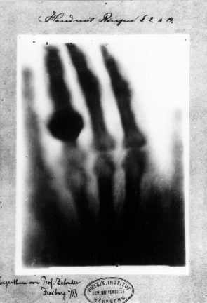
In the final days of 1895 a German physicist named Wilhelm Roentgen reported an intriguing discovery: the X-ray, a form of radiation that had enabled him to produce an image of the bones inside his wife Anna Bertha’s hand (as well as the wedding ring on her finger). When she first saw the image, an almost ghoulish rendering of her skeleton stripped of its skin, Anna Bertha herself said, “I have seen my death.” Until then, such a view would not have been possible until after her demise.
By thus opening up our interior selves for inspection, the discovery changed the ways we think of how we see. No longer was this confined to unobstructed views of people and things directly in front of our eyes. Now it also encompassed that which was previously inaccessible to us. Eventually, we even came up with a name for this new type of seeing: X-ray vision.
In fact, the idea of X-ray vision—in which the new technology would enable people to see things they hadn’t seen before, and maybe weren’t meant to see—emerged sooner, within weeks of Roentgen describing what he’d found. In its Feb. 1, 1896 issue, the British Medical Journal declared: “The application of the discovery to the photography of hidden structures is a feat sensational enough and likely to stimulate even the uneducated imagination.” The writer’s meaning here was clear: Quite aside from any medical value, the unwashed masses would want to know whether they could use the technology to see through people’s clothes.
To be sure, the discovery caused a bit of a panic among the more decorous elements of late-Victorian society. A London firm advertised “X-ray proof underclothing—especially for the sensitive woman,” and reportedly made a killing with it. Across the ocean, an assemblyman in New Jersey introduced a bill banning the use of X-rays in opera glasses.
The possibility of serving the more prurient interests wasn’t the only way X-rays stirred the public’s imagination, though. When Roentgen sat down with journalist H.J.W. Dam for an interview—the only interview he granted in the wake of the discovery—the first question Dam asked was, “Is the invisible visible?” The question referred, of course, to the newfound ability to peer inside the living body, to reveal its heretofore hidden frame, but underneath it lay another question, one with deeper, more profound implications: “Is the unknowable knowable?”
It wasn’t long before the themes of seeing and knowing and perhaps seeing and knowing too much were picked up by a proto-science fiction writer: the French author and Protestant minister Charles Recolin, who in 1896, only months after Roentgen’s discovery, published the story “Le Rayon X” (“The X-Ray”).
“Le Rayon X” tells the story of a Dr. Cornelius Schwanthaler, who injects one of his eyes with a serum, “an inoculable substance endowed with the astonishing power of making the eyes accessible to Roentgen rays—the famous rays then impassioning all Europe” (an English translation of the story is included in the 2012 collection The World Above The World, from Black Coat Press). His X-ray eye assists him admirably in the operating theater, but Schwanthaler is thinking beyond just surgery: “To see inside everything, to penetrate to the very center of matter, to scrutinize the framework that sustains the human membrane and perhaps discover, in those depths, the secret of the soul and the prime movement of thought was a double dream of medicine and philosophy.”
His X-ray vision proves too much, though. With the ability to see all, he is no longer capable of perceiving beauty, and illusion and hope are but bitter memories. Ecclesiastes was right, he decides: “Whoever increases knowledge increases sorrow. God has retained the worst part for himself: the truth.”
 |
|
The introduction of functional MRI led to studies probing what it is that makes us who we are
|
Nearly a century after the introduction of X-ray, the debut of the imaging technique functional magnetic resonance imaging reopened (albeit less dramatically than in the Recolin story) some of the same questions about the nature of seeing and knowing—casting an even keener eye, perhaps, on the matter of what makes us who we are. If X-ray made the invisible visible by revealing our inner anatomies—the structural constituents of our physical forms—fMRI delved deeper still, identifying and deconstructing the brain mechanisms underlying responses like fear and desire, which reside at the core of the human condition.
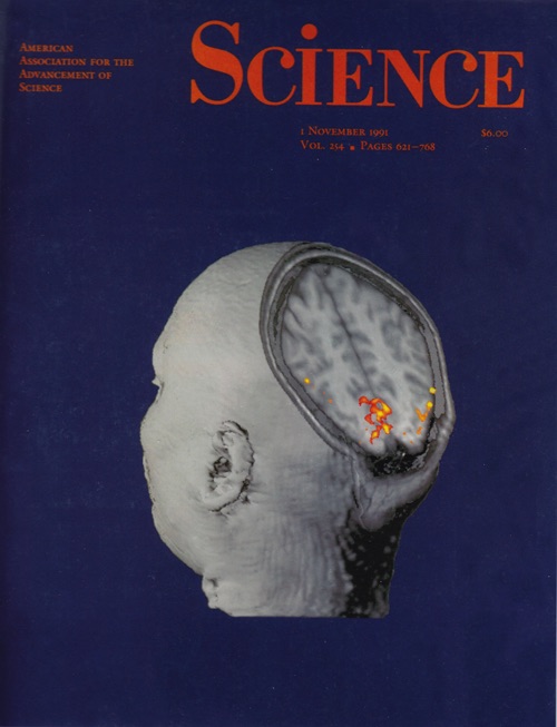
Functional MRI launched with a pair of papers by investigators in the Athinoula A. Martinos Center for Biomedical Imaging (then the MGH-NMR Center) at Massachusetts General Hospital in Boston. First was a November 1991 Science paper by Jack Belliveau and colleagues. Here, using dynamic susceptibility contrast MRI with a gadolinium-based contrast agent, Belliveau mapped the changes in cerebral blood volume following neural activation in a subject responding to a simple visual stimulus. His results represented the first images of human brain activity changes observed with MRI. The following June, in a paper published in Proceedings of the National Academy of Sciences, Kenneth Kwong reported a study in which he demonstrated MRI detection of activity in the brain based on endogenous contrast—contrast based on changes in the concentration of deoxyhemoglobin in the brain. Together, these studies ushered in the era of functional MRI and thus a revolution in the neuroimaging field.
Even then, in the very early days of fMRI, some envisioned using the technology to capture the essence of what it is to be human. Bruce Rosen, the Director of the Martinos Center, who played an instrumental role in the birth of the modality, describes one of the early motivations in developing fMRI: Belliveau, a brilliant mad scientist type if ever there was one, wanted nothing less than to be able to store human consciousness on a chip. “Jack worked on ‘brain mapping’ as a way to access all the memories and capabilities of the individual human brain,” Rosen says. “His fantasy goal was to use this data to fully encode a thinking brain, capturing all thoughts and actions in silico.”
The research community has yet to catch up with Belliveau’s original vision but it has nonetheless begun to circumscribe, in important ways, the mental processes underpinning human behavior. By measuring regional brain activity during cognitive tasks in healthy subjects, investigators have been able to associate particular cognitive functions with localized areas of the brain, and by extension explore how those areas work together in complicated neural networks to drive such behavior. In the quarter century since its introduction, functional MRI has helped to shed light on a broad range of higher-order cognitive functions, including learning and memory, attention, emotions, and even social cognition.
And there’s more. In a 2012 paper in the journal Neuroimage—“fMRI at 20: Has it changed the world?”—and in the talk on which the paper was based, the Lauterbur Lecture at the 2011 meeting of the International Society of Magnetic Resonance in Medicine, Rosen discussed three areas of inquiry that, collectively, address what might readily be described as “human nature” (or, to use the language of the Recolin story, “the soul”): consciousness, moral cognition and free will.
 |
|
In a lecture delivered to the International Society of Magnetic Resonance in Medicine, Bruce Rosen described fMRI studies of consciousness, moral cognition and free will
|
Functional MRI has expanded the conversation in each of these three domains. In the area of consciousness, researchers have upended long-held assumptions about vegetative and minimally conscious states, for example, by showing, in a patient in a vegetative state, the ability to understand spoken requests and to respond to them by imagining a particular task. Studies of moral cognition have demonstrated a complex and occasionally mutually competitive interplay of cognitive and emotional processes in making moral judgments. And finally, investigations of decision-making have raised thorny questions about free will. One study addressed head-on a controversy over whether “subjectively free” decisions are in fact predetermined by brain activity by showing a 10-second delay between encoding of a decision in the brain and the decision actually entering awareness.
Not surprisingly, perhaps, studies such as these have led to some amount of apprehension and disquiet over the potential impact of functional neuroimaging, even in the popular press. In 2002, for example, The Economist published a cover story on the ethics of brain science that opened with a foreboding observation: “Genetics may yet threaten privacy, kill autonomy, make society homogeneous and gut the concept of human nature. But neuroscience could do all of these things first.” And primary among the possible instruments of bringing about this not-so-distant dystopian future? Yes, fMRI.
The article was especially concerned with how neuroimaging was disrupting our notion of free will. Not least of what modern-day philosophers were wrestling with: “the idea that mental decisions are purely the consequence of electrochemical interactions in the brain [and] the separate, but parallel, argument that correct moral choices are the result of a sort of biological decision-making programme, shaped by evolution, rather than being arrived at by abstract reasoning.”
And existential tumult hasn’t been the only distressing byproduct of the new imaging modality. As in the wake of the discovery of the X-ray, the development of fMRI has given rise to concerns about more sordid possible uses—the 21st-century version, perhaps, of wanting to see through people’s clothes. Rosen points to the burgeoning field of “neuromarketing” as an example. Neuromarketing, which considers consumers’ cognitive and affective responses to marketing stimuli in developing marketing messages, might seem essentially the same as conventional marketing—both seek to tease out consumers’ unstated needs and desires—but to many the idea of using neuroimaging to achieve this is somehow unsettling. “Perhaps the key difference here is that their decisions are based on information inaccessible even to ourselves,” Rosen says.
Still, even given these various concerns, functional MRI must be viewed as having served us remarkably well over the past 25 years. Not only has it contributed to better clinical care, the technology has helped us see and understand ourselves in new and important ways.
“There will always be potential for abuse, in areas like neuromarketing, lie detection and other so-called ‘mind-reading’ applications,” Rosen says, “but the advancement of fMRI has been, and will remain, an undeniably positive development in the history of biomedical imaging. This is true both clinically and in society at large. The technology has contributed to pre-surgical mapping for neurosurgery applications, improved the diagnosis and care of patients with a range of psychiatric diseases and disorders, and ultimately given us a more enlightened view of fundamental aspects of human nature."


