Currently, our lab is active in a number of research projects related to brain tumors, hypoxic ischemic injury in neonates, neurodegenerative diseases including amyotrophic lateral sclerosis (ALS), and neuro-developmental disorders such as autism or OCD
The goals of our lab are to advance the understanding of central nervous system diseases through the application of advanced neuroimaging techniques, specifically, magnetic resonance imaging (MRI) and MR Spectroscopy (MRS).
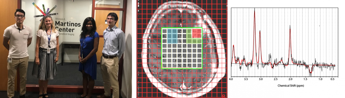
Magnetic resonance spectroscopy (MRS) is a magnetic resonance-based imaging modality that allows noninvasive sampling of metabolic changes in normal and abnormal brain parenchyma. MRS is particularly useful in the differentiation of developmental or non-neoplastic disorders from neoplastic processes. MRS is also useful during routine imaging follow-up after radiation treatment or during antiangiogenic treatment and for predicting outcomes and treatment response.
Proton magnetic resonance spectroscopy (1H MRS) is a noninvasive imaging technique that can easily be added to the conventional magnetic resonance (MR) imaging sequences. Using MRS one can directly compare spectra from pathologic or abnormal tissue and normal tissue. Metabolic changes arising from pathology that can be visualized by MRS may not be apparent from anatomy that can be visualized by conventional MR imaging. In addition, metabolic changes may precede anatomic changes.
OBJECTIVE: Investigating consequences of early or late antiretroviral therapy (ART) initiation in infancy on young brain development using magnetic resonance spectroscopy.
DESIGN: Most pediatric HIV/ART-related neurological studies are from neuropsychological/clinical perspectives. Magnetic resonance spectroscopy can elucidate the mechanisms underpinning neurocognitive outcomes by quantifying the brain's chemical condition through localized metabolism to provide insights into health and development.
Sphingosine 1-phosphate (SP1) receptors may be attractive targets for modulation of inflammatory processes in neurodegenerative diseases. Recently fingolimod, a functional S1P1 receptor antagonist, was introduced for treatment of multiple sclerosis. We postulated that anti-inflammatory mechanisms of fingolimod might also be protective in Alzheimer's disease (AD).
PURPOSE: Measurements of objective response rates are critical to evaluate new glioma therapies. The hallmark metabolic alteration in gliomas with mutant isocitrate dehydrogenase (IDH) is the overproduction of oncometabolite 2-hydroxyglutarate (2HG), which plays a key role in malignant transformation. 2HG represents an ideal biomarker to probe treatment response in IDH-mutant glioma patients, and we hypothesized a decrease in 2HG levels would be measureable by in vivo magnetic resonance spectroscopy (MRS) as a result of antitumor therapy.