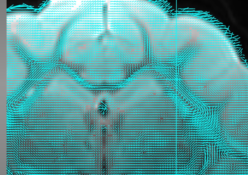
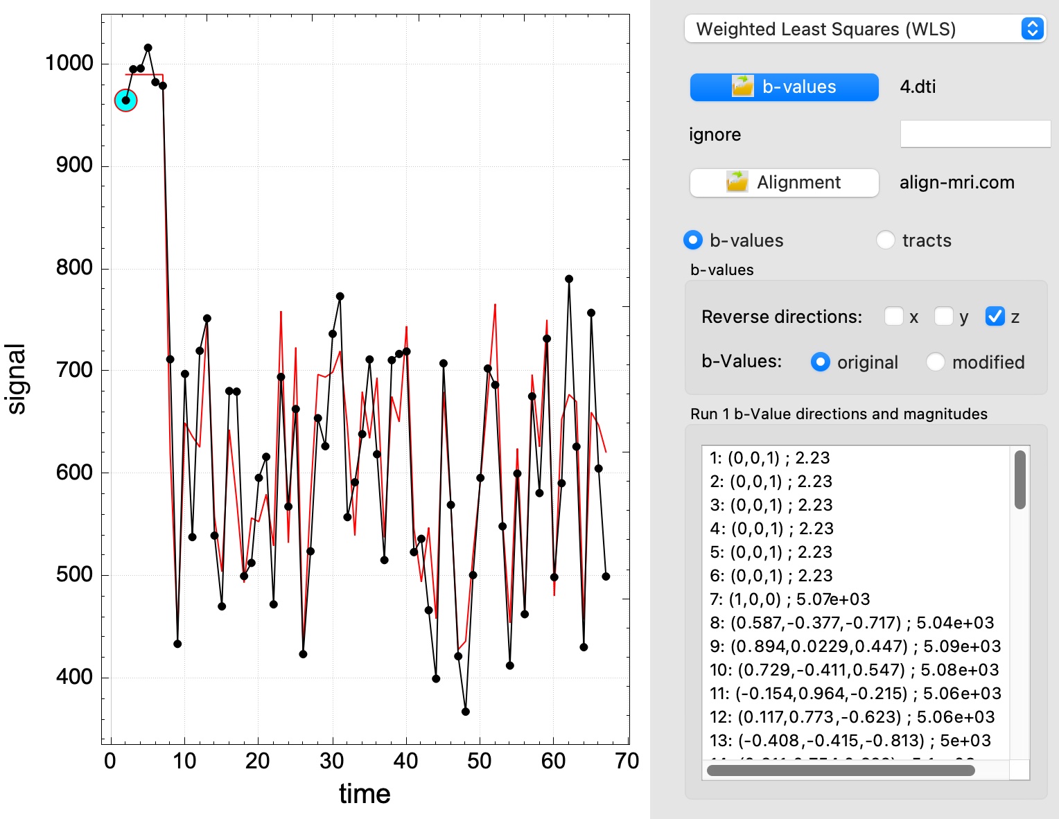
Diffusion tensor images can be fit using either ordinary or weighted least squares and the conventional diffusion tensor model. Diffusion b-values and directions are read from a table for each run, with 4 entries per time point specifying the diffusion unit vector and magnitude (b_x, b_y, b_z, b_mag). Directions can be reversed if necessary to account for mis-registration of subjects in the scanner or other directional problems. If data have been spatially rotated for registration (“normalization”) using fastmap, then the alignment parameters can be used to rotate the b-vectors from the scanner frame to the rotated presentation. Saved maps include
- Fraction anisotropy (FA) as a vector value and as a scalar
- Mean diffusivity (MD)
- Longitudinal diffusivity (LD)
- Radial diffusivity (RD)
- Signal at b=0 (S0)
To enable tract tracing by tensor deflection, tensor maps are saved as 8 components: Dxx, Dyy, Dzz, Dxy, Dxz, Dyz, SEM(diagonal), SEM(off-diagonal)
Tract tracing is accomplished by tensor deflection with an initial step in which a tract is traced from every voxel within a specified restriction region (e.g., brain), followed by a second step in which one or more tracts are traced that intersect with specified voxels or regions of interest.